
Welcome to ProLine Sports Nutrition
ProLine Sports Nutrition is owned and managed by endurance athletes. While training, we searched for the best supplements to enhance our performance without compromising our ability to compete against world class athletes in professional and amateur sporting leagues.

First Endurance Optygen HP
OptygenHP is a legal, safe and stimulant-free formula that's engineered to increase endurance capacity.
Featured Products
-
, BPN Endo Pump Pre-Workout Muscle Pump Enhancer, Increased Blood Flow/Oxygen Transport to Muscles, Blackberry Lemonade
Vendor:PROLINE SPORTS NUTRITIONRegular price $47.31 USDRegular priceUnit price per$0.00 USDSale price $47.31 USDSold out -
, BPN Flight Pre Workout, Blue Raspberry, 30 Servings
Vendor:PROLINE SPORTS NUTRITIONRegular price $55.22 USDRegular priceUnit price per$0.00 USDSale price $55.22 USD -
, BPN G.1.M Go One More Sport, Endurance Training Fuel, Electrolytes and Calories, Pink Himalayan Salt, Salted Watermelon
Vendor:PROLINE SPORTS NUTRITIONRegular price $49.78 USDRegular priceUnit price per$0.00 USDSale price $49.78 USD -
, BPN Strong Greens Superfood Powder, Improved Digestion, Increased Energy, Immune System Support, Lemon
Vendor:PROLINE SPORTS NUTRITIONRegular price $55.97 USDRegular priceUnit price per$0.00 USDSale price $55.97 USD


Ride with the best.
SportLegs Stops Muscle Burn capsules are the ideal accompaniment to an active lifestyle. Take your active lifestyle to the next level with SportLegs Stops Muscle Burn capsules! Designed specifically for active individuals, these capsules will help you push through muscle burn and reach your ultimate potential. Don't let soreness hold you back any longer - try SportLegs today!


GU Energy
GU Energy Original Sports Nutrition Energy Gel, 8 Count Pack


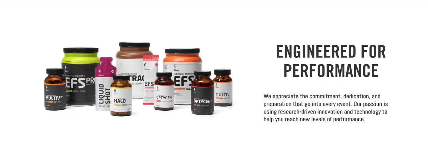
First Endurance Premium Nutrition
First Endurance
OptygenHP, OPTYGEN, now with OptyMax- The Next Level for Cyclists,
Triathletes and Elite Endurance Athletes. Read reviews and learn from
the professionals.
First Endurance Optygen HP with Ashwagandha Root the most potent form now available for your season is
designed to help you optimize performance, maximize oxygen utilization
and achieve greatness. The patent-pending, legal, safe and
stimulant-free Optygen formula
is based on human clinical trials and the latest scientific research on
increasing endurance. It's so effective, there's a 100% money back
performance guarantee. First Endurance Optygen is used my many endurance athletes who want to take their game to the next level. First Endurance Optygen
Read our Official Athlete Review of First Endurance Optygen HP
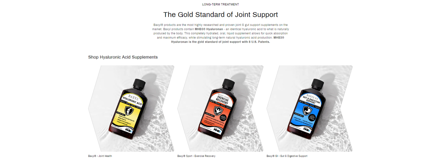
Performance and Recovery
-
Premium Endurance Supplements
Discover MoreEndurance supplements are products to help athletes and individuals improve their physical performance and stamina during exercise. They come in various forms, including powders, capsules, gels, and bars.
-
Premium Recovery Supplements
Recover FasterMuscle recovery supplements are products to aid the body's natural process of repairing and rebuilding muscle tissue after exercise. They come in various forms, including powders, capsules, gels, and bars.
-
Specialty Sports Supplements
Learn MoreSpecialty sports supplements are a category of supplements that cater to specific needs of athletes and individuals engaged in intense physical activities. These supplements go beyond the basic protein powders and BCAAs, targeting particular aspects of performance and recovery
Featured Products
-
VO2-BOOST Endurance Supplement 120 Capsules
Regular price $44.95 USDRegular priceUnit price per$59.95 USDSale price $44.95 USDSale -
EPO-BOOST BodyEndurance Complex 120 Capsules
Regular price $44.95 USDRegular priceUnit price per$59.95 USDSale price $44.95 USDSold out -
OptygenHP Premium Endurance Supplement 120 Capsules | First Endurance Optygen HP
Regular price $79.95 USDRegular priceUnit price per -
HAMMER NUTRITION Heed, Mandarin-Orange 32 servings Classic 2.0 (32 Servings)
Regular price $37.95 USDRegular priceUnit price per


